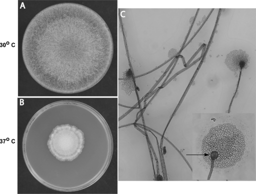
FIG. 1.
Rhizomucor variabilis var. regularior. (A and B) Macroscopic morphology on potato dextrose agar. Colonies were filling the entire plate in 5 days at 30°C (A) and showed restricted growth at 37°C (B). Colonies varied in color from brown to tan and were hairy, with reverse buff-to-brown color. (C) Microscopic morphology in lactophenol cotton blue stain after 3 days (magnification, ×200): hyaline, unbranched, ribbon-like hyphae; long, simple sporangiophores arising from hyphae and ending in sporangium; spherical sporangia with globose columella and no apophysis; hyaline, ellipsoidal, smooth-walled sporangiospores. The inset shows details of the spherical columella (arrow) with sporangiospores (magnification, ×400).

