FIG. 5.
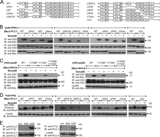
Analysis of PLCγ domains required for plexin-B1-mediated signaling. (A) Graphical representation of the analyzed PLCγ1 mutants. (B and D) HEK 293 cells were transfected with plexin-B1, PDZ-RhoGEF (PRG), wild-type PLCγ1 (WT), or its mutants. Cells were incubated without (−) or with (+) 150 nM Sema4D and lysed, and the interaction between plexin-B1 and PLCγ1 and its mutants was analyzed by immunoprecipitation (B), or the amount of activated RhoA was analyzed as described in Materials and Methods (D). (C) Wild-type or Y/F mutants of plexin were cotransfected with the Myc-tagged N- or C-terminal SH2 domain of PLCγ1 (SH2.N or SH2.C, respectively). Protein-protein interactions were analyzed by coimmunoprecipitation. (E) MCF-7 cells were treated with phosphate-buffered saline (ctrl.), 150 nM Sema4D, or 10 ng/ml EGF. Cells were lysed, and immunoprecipitation (IP) was performed using anti-PLCγ1 or antiphosphotyrosine antibodies (left and right panels, respectively). Samples were then analyzed by immunoblotting (IB) using the indicated antibodies.

