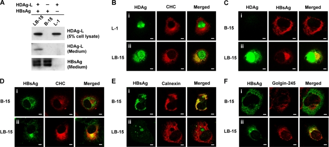
FIG. 6.
Subcellular localizations of HDAg-L, small HBsAg, and CHC in stable cell lines. (A) Western blot analysis. HDAg-L and small HBsAg in stable cell lines L1, B-15, and LB-15 and the culture media were examined by Western blot analysis. (B to F) Immunofluorescence staining. Subcellular localizations of HDAg-L, small HBsAg, and CHC in the stable cell lines were examined by immunofluorescence staining with antibodies against HDAg, CHC, and organelle markers calnexin and Golgin-245 using confocal microscopy. Bars, 20 μm.

