FIG. 1.
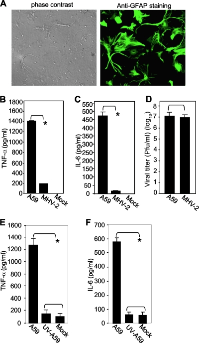
Differential induction of TNF-α and IL-6 in primary mouse astrocytes by infection with MHV-A59 and MHV-2. (A) (Right) The purity of isolated primary mouse astrocytes was determined by immunofluorescence staining with an antibody specific for GFAP. (Left) Phase-contrast image showing the same field as in fluorescence staining. (B and C) Induction of TNF-α (B) and IL-6 (C) in primary astrocytes by MHV infection. Cells were infected with MHV-A59 or MHV-2 at an MOI of 5 or mock infected as a control. The culture supernatants were collected at 24 h p.i. The amounts of TNF-α (B) and IL-6 (C) proteins in the supernatants were determined using ELISA kits. The results are expressed as the mean number of picograms per milliliter for three independent experiments. The error bars indicate standard deviations of the means. The asterisks indicate the statistical significance between the pairs (P < 0.05). (D) Virus production in primary mouse astrocytes. Virus infection was carried out as for panels B and C, and virus titers were determined at 24 h p.i. by plaque assay on DBT cells. The results are expressed as the mean number of PFU per milliliter for three independent experiments. The error bars indicate standard deviations of the means. (E and F) Dependence of TNF-α and IL-6 induction on virus replication. Primary astrocytes were infected with live or UV-inactivated MHV-A59 at an MOI of 5 or mock infected. At 24 h p.i., the culture supernatants were collected to determine TNF-α (E) or IL-6 (F) protein amounts by ELISA as for panels B and C.

