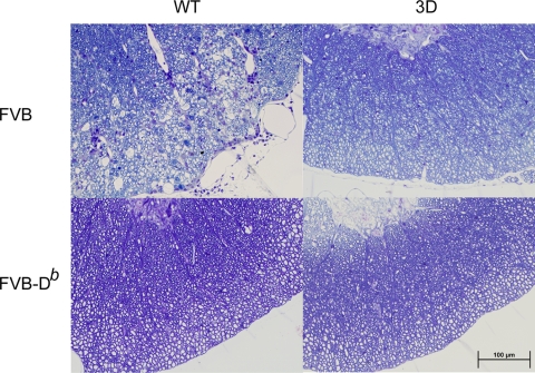
FIG. 5.
Spinal cord sections of mice at 90 dpi. Plastic-embedded sections were stained with a modified erichrome-cresyl violet stain. Lateral columns of cord show inflammation and demyelination in FVB mice but not in FVB-3D, FVB-Db, or FVB-Db.3D mice. All pictures were taken in a similar region of the spinal cord with a ×40 objective. WT, wild type.

