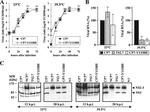
FIG. 4.
Characterization of BVDV CP7-Y1338H. (A) Growth kinetics of BVDV CP7 and the genetically engineered ts mutant CP7-Y1338H at 33°C (left) and 39.5°C (right). MDBK cells were infected at an MOI of 0.1 with supernatants of the second cell culture passages of the indicated viruses. The titers of released virus were determined by IF analysis over a time period of 72 h. Error bars indicate the ± ranges from quadruplicate titrations. (B) Relative amounts of accumulated viral genomic RNAs obtained after infection of MDBK cells with BVDV CP7, TS2.7, and CP7-Y1338H at 33°C (left) and 39.5°C (right). For infection an MOI of 1 was used. Total cellular RNAs were extracted at 10 h p.i. and subjected to quantitative BVDV-specific real-time RT-PCR. Results are indicated as percentages of the mean value obtained for reference strain CP7 (100%). Data show the means ± standard deviation ranges from two independent experiments, each analyzed by the measurement of triplicates. (C) Immunoblot. MDBK cells infected with CP7, NCP7, TS2.7, and CP7-Y1338H at 33°C (left side) and 39.5°C (right side) were lysed 12 and 20 h p.i. (h.p.i.). For infection an MOI of 2 was used. The samples were separated by sodium dodecyl sulfate-polyacrylamide gel electrophoresis (8% polyacrylamide) under reducing conditions, transferred to nitrocellulose, and incubated with the anti-NS3 monoclonal antibody 8.12.7 (15). Noninfected (n.i.) cells served as negative controls. The sizes (in kDa) of the molecular mass marker proteins (in thousands) are indicated on the left. The positions of NS2-3 and NS3 are indicated on the right.

