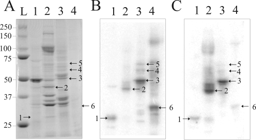
FIG. 1.
Detection of Prunus persica proteins interacting with PLMVd by Northwestern blot analysis. (A) Coomassie blue staining of the 10% PAGE gel. (B and C) Proteins were transferred onto nitrocellulose membranes, which were then probed using the (−) (B) and (+) (C) PLMVd strands. The blots show total protein extract from peach tree leaves (lanes 1) and purified protein fractions obtained after fractionation on a heparin-Sepharose column and subsequent elution with 150 mM (lanes 2), 400 mM (lanes 3), and 600 mM (lanes 4) ammonium sulfate. In panel A, lane L is a molecular weight marker. The numbering of the bands corresponds to that used in Table 1.

