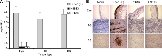
FIG. 1.
(A) Viral replication in the eye, trigeminal ganglia, and brain. Groups of 6-week-old female BALB/c mice were mock infected or infected with HSV-1(F), R3616, or H9813 at 4 × 10⁵ PFU through corneal scarification. At 5 days postinfection, eye, trigeminal ganglia (TG), and brain stem (BS) tissues were collected to determine virus yields. Data are expressed as means ± standard deviations for six mice for each group. (B) Immunohistochemistry staining of mouse tissues. The sections from eye, trigeminal ganglia, and brain stem tissues described above were reacted with anti-HSV-1 antibody, and immunohistochemistry was performed. Specific HSV-1 staining is shown in brown. Representative images from each mouse group were chosen for the panels.

