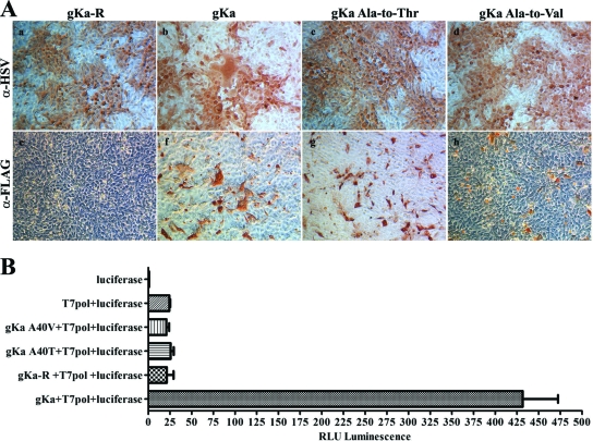
FIG. 8.
Ability of the gKa peptide to complement gKΔ31-68/gBΔ28syn-induced cell fusion. (A) Vero cells were transfected with the gKa, gKa-R, gKa-A40V, or gKa-A40T plasmid, and cells were subsequently infected with the gKΔ31-68/gBΔ28syn virus at an MOI of 0.2. Cells were reacted with the anti-FLAG (α-FLAG) or the anti-HSV (α-HSV) antibody under methanol-fixed conditions and visualized by phase-contrast microscopy after immunostaining. (B) Vero cells were transfected and infected as detailed above, with the exception that two different populations of cells were transfected at the same time, with each of the gK-expressing plasmids with a plasmid expressing T7 polymerase (pol) or a plasmid expressing the luciferase gene under the T7 promoter. These equal populations of cells were mixed together prior to infection with the gKΔ31-68/gBΔ28syn virus. The amount of relative fusion was obtained by measuring the relative level of luminescence (RLU) emitted by cellular extracts at 24 hpi. Error bars indicate the standard error.

