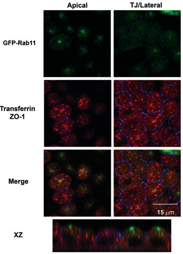
Figure 1. Distribution of recycling endosomes in polarized MDCK cells.
Filter-grown MDCK cells stably expressing GFP-Rab11 (green) were incubated with basolaterally added canine Tf for 30 min, then fixed and processed for indirect immunofluorescence with antibodies against canine Tf (in red) and the tight junction marker ZO-1 (in blue). Individual and merged confocal sections taken just beneath the apical surface and at the level of the tight junction/lateral border are shown. An xz section is shown in the bottom panel. Note the segregation of Rab11 and transferrin, which mark the apical recycling and common recycling endosomes, respectively.

