
Figure 7.
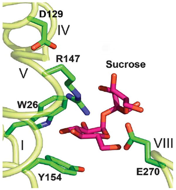
Sucrose molecule docked into the putative sugar-binding site of the CscB model. The sucrose molecule (coordinates from PDB, ID 1IW0), is modeled in the putative sugar-binding site of CscB using the program XtalView.45 Essential amino acid residues are shown as sticks. Transmembrane helices are numbered in roman numerals.