Figure 1.
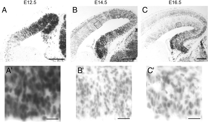
β-Catenin signaling levels undergo progressive reduction during cortical neurogenesis. Comparable coronal sections from E12.5 (A), E14.5 (B), and E16.5 (C) BAT-gal mouse cortex after X-gal staining reaction. BAT-gal mice express β-galactosidase in response to β-catenin signaling. Additional detail of the ventricular zone is shown in high-power insets below (A′, B′, C′). Scale bars: low-power images, 100 μm; high-power images, 20 μm.

