Figure 7.
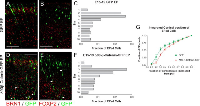
Alteration of β-catenin signaling during late neurogenesis influences cell positioning and fate. A–F, E15.5 embryos were electroporated with GFP (A–C) (n = 4 brains) or Δ90β-catenin-GFP (D–F) (n = 4 brains) constructs. Embryos were analyzed at E19.5 and sections were stained for GFP and BRN1, a marker for cortical layers 2–4 (A, D) or FOXP2, a marker for cortical layers 5 and 6 (B, E). The fractions of GFP+ cells that coexpressed BRN1 were significantly different (p = 0.0022, t test, two tailed). Increasing β-catenin signaling by Δ90β-catenin-GFP decreased the fraction of BRN1+ cells (0.8983 ± 0.01558, n = 206) when compared to control (0.9785 ± 0.002237, n = 247). The fractions of GFP+ cells coexpressing FOXP2 were also significantly different (p = 0.0397, t test, two tailed). Increasing β-catenin signaling by Δ90β-catenin-GFP increased the fraction of FOXP2+ cells (0.07780 ± 0.01518, n = 363) when compared to control (0.02693 ± 0.01214, n = 148). The somal position of each electroporated cell within 10 equally sized bins covering the complete cortical plate was also noted. The fraction of the total GFP+ cells in each of the 10 bins was then graphed (C, F) for both experimental conditions. Bin 1 corresponds to the section of the cortical plate closest to the pial surface, while bin 10 is adjacent to the white matter tracts. Overall, cells electroporated with Δ90β-catenin-GFP (n = 447) resided in slightly deeper cortical positions than GFP (n = 280) control cells. G, When these results are shown in a cumulative view, the shift in positioning is more evident. Arrows highlight examples of FOXP2/GFP double-positive cells. Arrowheads indicate GFP+/BRN1− cells. Scale bars, 100 μm. EPed, Electroporated.

