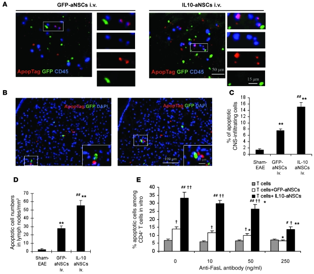
Figure 6. Enhanced proapoptotic effect of IL-10–aNSCs on inflammatory cells.
Mice treated with aNSCs i.v. at day 22 p.i. were sacrificed 2 weeks p.t. Brains, spinal cords, and lymph nodes were harvested. The same region of the corpus callosum was examined in all groups, as shown in Supplemental Figure 3. (A) Colocalization of apoptotic cells (red) and CD45+ (blue) in the brain indicated that the majority of apoptotic cells were CNS-infiltrating cells, while no GFP+ aNSCs (green) underwent apoptosis. (B) GFP+ aNSCs were present in lymph node sections, none of which underwent apoptosis. Nuclei in B were stained with DAPI (blue). The insets show high-magnification images of the boxed regions. Original magnification, ×20 (A); ×10 (B), ×65 (insets, A and B). Quantitative analysis of apoptotic CNS-infiltrating cells (C) and lymph node cells (D). Symbols represent mean ± SD; n = 6–8 mice per group. **P < 0.01, comparisons between sham-EAE group and other groups; ##P < 0.01, comparison between GFP-aNSCs i.v. and IL-10–aNSCs i.v. (E) Block of FasL inhibited the proapoptotic effect of aNSCs on T cells in cocultures. Apoptotic cells were defined as CD4+, Annexin-V+, and DAPI– by flow cytometry. Results are mean ± SEM of 4 independent experiments. #P < 0.05, ##P < 0.01, comparison between T cells cocultured with GFP-aNSCs and with IL-10–aNSCs. †P < 0.05, ††P < 0.01, comparison between T cells cocultured with aNSCs and T cells alone. *P < 0.05, **P < 0.01, comparison between wells unblocked and blocked by anti-FasL antibody.

