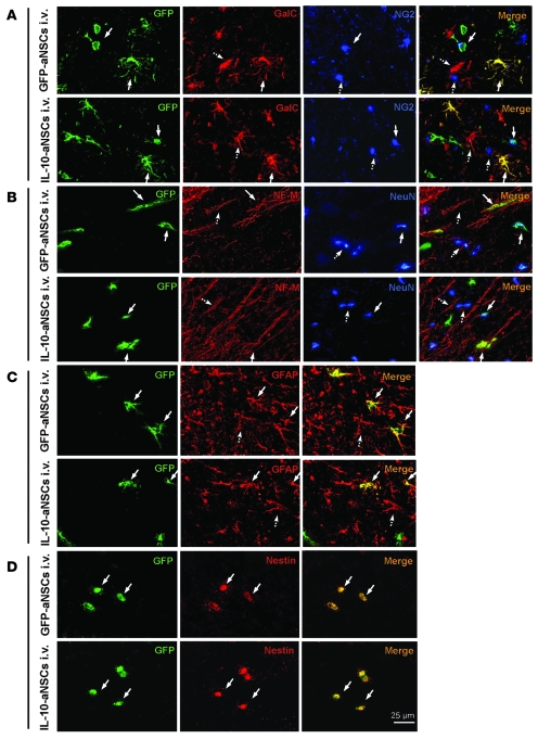
Figure 9. IL-10–aNSCs selectively expand neuron and oligodendrocyte populations in vivo.
Mice treated with aNSCs i.v. at day 22 p.i. were sacrificed at day 78 p.t., and brains were harvested for immunohistology. The same region of the corpus callosum was examined in all groups, as shown in Supplemental Figure 3. (A–D) Immunofluorescence images of the brain in aNSC-treated mice at day 78 p.t. Cells colabeled with GFP (green) and neural-specific markers (red, blue) were identified as differentiated cells derived from transplanted aNSCs (arrows with solid lines), which were morphologically indistinguishable from respective endogenous cells (arrows with dashed lines). Some of the transplanted aNSCs remained nestin+ (undifferentiated). Original magnification, ×40 (A–D).

