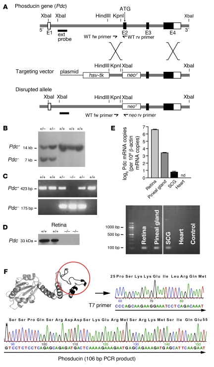
Figure 1. Generation of mice deficient in Pdc.
(A) Targeting vector for disruption of Pdc in mouse ES cells via homologous recombination. Insertion of a neomycin resistance gene (neor) disrupted the exon 2 (E2) of the Pdc gene including the start codon. Fw, forward primer; rv, reverse primer; ATG, position of the start codon of the Pdc gene. (B) Southern blot analysis of E14 mouse ES cells transfected with the Pdc targeting vector. Using a 5′ external probe, the wild-type allele (+/+) was identified as a 14-kb band, whereas the targeted allele (+/–) was detected as a 7-kb signal (after incubation of genomic DNA with XbaI). (C) Identification of wild-type or deleted Pdc alleles (+/–, –/–) by PCR analysis of genomic DNA obtained from tail biopsies of mice. (D) Absence of Pdc protein in retina of Pdc–/– mice as confirmed by Western blotting with a polyclonal Pdc antiserum. (E) Top: Quantitative PCR analysis of Pdc mRNA levels in wild-type retina, pineal gland, SCG, and cardiac ventricles (Pdc mRNA copy number normalized to 106 Actb mRNA copies, n = 3–5 per group). Bottom: PCR reactions in the plateau phase were loaded on a 2% agarose gel to verify PCR product size (106 bp) (lanes were run on the same gel but were noncontiguous). nd, not detectable. (F) The identity of the PCR products from SCG was verified by subcloning and sequencing, which identified a sequence corresponding to amino acids 25–55 in Pdc (circle, black ribbon part in Pdc structure).

