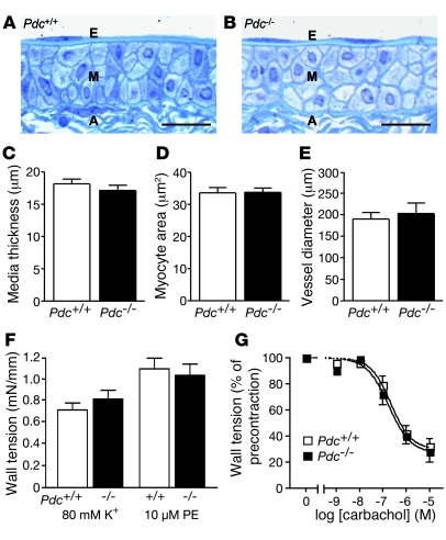
Figure 3. Vascular function in Pdc-deficient mice at a young age.
(A–D) Longitudinal sections through the iliac artery of wild-type (A) and Pdc–/– mice (B) revealed no alterations in microscopic structure, media thickness (C), or vascular smooth muscle cell cross-sectional area (D) in Pdc–/– mice (n = 4 per genotype, age 1.5–2 months). Scale bars: 20 μm. E, endothelium; M media layer; A, adventitia. (E) Internal diameter of isolated iliac artery segments mounted in a small vessel myograph and prestretched to a wall tension corresponding to 100 mmHg intraluminal pressure (n = 6 per genotype). (F) Vasoconstrictory response to depolarization by 80 mM K+ or α1 adrenoceptor activation by phenylephrine was similar in Pdc–/– and Pdc+/+ iliac artery segments (n = 6–10). (G) Vasorelaxation induced by the muscarinic receptor agonist carbachol was unaltered in Pdc–/– compared with Pdc+/+ vessels. Vessel segments were precontracted by 10 μM phenylephrine (n = 6 vessels per genotype).

