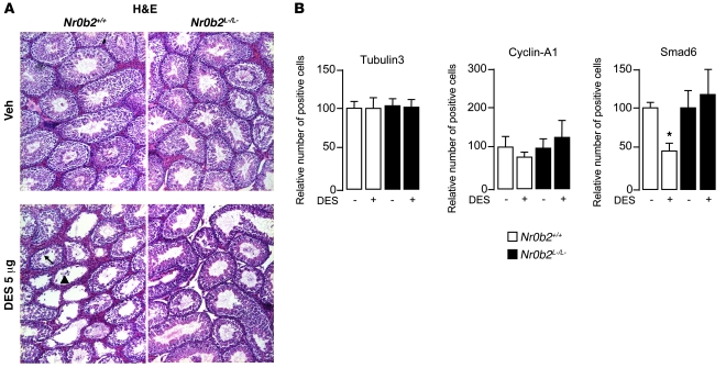
Figure 2. DES-induced histological abnormalities caused by loss of postmeiotic cells in Nr0b2+/+ mice.
(A) Representative micrographs of H&E-stained testes of 10-week-old Nr0b2+/+ and Nr0b2L–/L– mice exposed to vehicle or 5 μg DES (n = 6 per group). The arrow indicates tubules with a slight loss of germ cells; the arrowhead indicates tubes with complete loss of germ cells. Original magnification, ×100. (B) Quantification of cells per 100 seminiferous tubules (n = 4–6) positively stained for markers Smad6 (postmeiotic germ cells), Tubulin3 (Sertoli cells), and Cyclin-A1 (pre- and meiotic germ cells). Vehicle-treated mice were arbitrarily set at 100%. *P < 0.05 versus vehicle.

