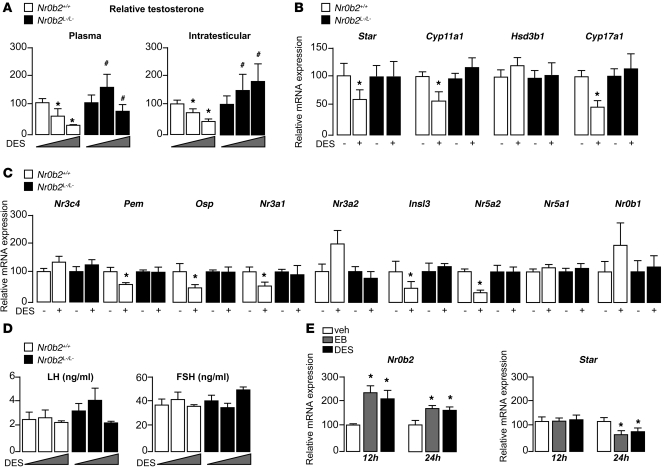
Figure 4. Nr0b2 controls DES-induced repression of testosterone synthesis.
(A) Plasma and intratesticular testosterone levels in 10-week-old Nr0b2+/+ and Nr0b2L–/L– mice exposed to 0, 0.75, or 5 μg DES (n = 10–15 per group). (B) Testicular mRNA expression of Star, Cyp11a1, Hsd3b1, and Cyp17a1, normalized to β-actin levels, in whole testes of 10-week-old Nr0b2+/+ and Nr0b2L–/L– mice exposed to 0 or 0.75 μg DES (n = 10–15 per group). (C) Testicular mRNA expression of Nr3c4, Pem, Osp, Nr3a1, Nr3a2, Insl3, Nr5a2, Nr5a1, and Nr0b1, normalized to β-actin levels, in whole testes of 10-week-old Nr0b2+/+ and Nr0b2L–/L– mice exposed to 0 or 0.75 μg DES (n = 10–15 per group). (D) Plasma LH and FSH concentration in 10-week-old Nr0b2+/+ and Nr0b2L–/L– mice exposed to 0, 0.75, or 5 μg DES (n = 10–15 per group). (E) mRNA expression of Nr0b2 and Star normalized to β-actin levels in MA-10 Leydig cells exposed to vehicle, EB, or DES for 12 or 24 hours (n = 6 per group). Vehicle-treated mice were arbitrarily fixed at 100%. *P < 0.05 versus vehicle; #P < 0.05 versus Nr0b2+/+ given the same DES dose.

