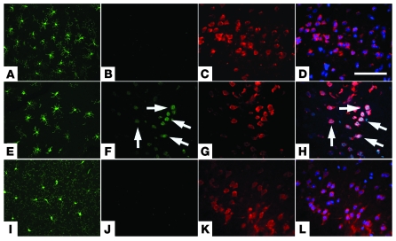
Figure 2. LPS administration provokes neuroinflammation and neuronal CCEs.
(A–D) Nontransgenic mice at 2 months of age subject to LPS injections exhibited Iba1-immunoreactive neocortical microglia with an activated morphology (A) with no evidence of expression of cyclin D (B) in NeuN-positive neurons (C). (E–H) Age-matched R1.40 transgenic animals injected with LPS exhibited Iba1-positive microglia with an activated morphology (E) as well as expression of cyclin D (F) in a subset of NeuN-positive cortical layer II/III neurons (G). (I–K) Age-matched R1.40 animals injected with PBS exhibited Iba1-positive microglia with a resting morphology (I) and no evidence of expression of cyclin D (J) in NeuN-positive neurons (K). Similar results were obtained with immunohistochemistry for the cell cycle protein cyclin A (not shown). (D, H, and L) Merged images. Nuclei were counterstained with DAPI (blue). Arrows indicate cyclin D–positive neurons. Scale bar: 100 μm.

