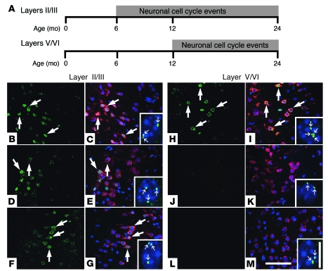
Figure 7. Therapeutic trial of NSAIDs inhibits subsequent, but not extant, neuronal CCEs.
(A) Neuronal CCEs were first observed in frontal cortical layers II/III at 6 months of age and persisted for 2 or more years in the R1.40 animals. Neuronal CCEs were not observed in deeper cortical layers V/VI until 12 months of age. (B–M) R1.40 transgenic mice at 6 months of age were fed control (B, C, H, and I), ibuprofen-containing (D, E, J, and K) or naproxen-containing (F, G, L, and M) diets for 6 months. (B and H) Control diet–fed mice exhibited expression of cyclin D (large arrows) in NeuN-positive neurons in frontal cortex layers II/III and layers V/VI. (D, F, J, and L) Ibuprofen- or naproxen-containing diet–fed mice exhibited expression of cyclin D in a subset of NeuN-positive neurons in layers II/III (D and F), with minimal expression of cyclin D in layers V/VI (J and L). (C, E, G, I, K, and M) Merged images. Sections were stained with NeuN (red), and nuclei were counterstained with DAPI (blue). FISH analysis with a DNA probe specific for mouse chromosome 16 demonstrated the presence of a subset of neuronal nuclei with 3 or 4 spots of hybridization (small arrows) in all treatment groups in cortical layers II/III (C, E, and G, insets) and only the control diet group in cortical layers V/VI (I, K, and M, insets). Scale bars: 100 μm (B–M); 10 μm (insets).

