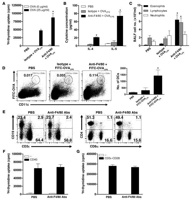
Figure 7. IMs prevent LPS-triggered Th2 responses to innocuous inhaled antigens.
(A–G) Naive BALB/c mice were injected i.p. daily from day 1 to 3 with depleting anti-F4/80 or control isotype antibodies. (A–C) On day 2, mice received an i.t. injection of OVALPS. (A and B) On day 6, MLN cells were restimulated for 3 days with 25 μg/ml OVA. The proliferation was measured (A), and culture supernatants were assayed for IL-4 and IL-5 by ELISA (B). (C) From day 11 to 14, mice were challenged intranasally with 25 μg OVA (grade V; Sigma-Aldrich) in 50 μl PBS. On day 15, BALF was subjected to differential cell counts. (D) On day 2, IM-depleted and control mice were injected i.t. with 100 μg FITC-OVA (rather than unlabeled OVA) and 10 ng LPS. On day 3, MLNs were analyzed by flow cytometry for the presence of OVA-loaded DCs (FITC+F4/80–CD11c+). Percentages (left) and total numbers (right) of migrating DCs are shown. (E) On day 4, the percentages of T (CD3ε+, CD4+, or CD8α+ cells) and B (CD19+ cells) cells in MLNs were measured by flow cytometry. (F and G) On day 4, B and T cells were isolated from MLNs and stimulated ex vivo with agonist anti-CD40 antibodies or anti-CD3 and anti-CD28 antibodies, respectively. Control cells were left unstimulated. B cell (F) and T cell (G) proliferation was measured as [3H]thymidine incorporation during the last 16 hours of a 2-day culture. *P < 0.05 versus results obtained with isotype control antibodies (A–D).

