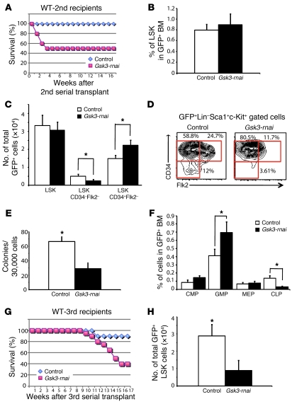
Figure 4. Gsk3 knockdown depletes HSCs in serial BM transplants.
(A) Noncompetitive serial transplants were performed by transplanting 2 × 10⁵ sorted GFP+ cells from primary recipients of control or Gsk3-rnai transduced BM into lethally irradiated recipients (10 mice per group). Survival of secondary recipients receiving control or Gsk3-rnai BM is shown as a Kaplan-Meier plot. (B) Percent HSC-containing LSK fraction in control and Gsk3-rnai secondary recipients. (C) Absolute number of GFP+ LSK, LSK CD34–Flk2–, and LSK CD34+Flk2– cells in control and Gsk3-rnai secondary recipients. (D) Representative FCM data, presented as the distribution of CD34–Flk2–, CD34+Flk2–, and CD34+Flk2–, which immunophenotypically correspond to LT-HSCs, ST-HSCs, and MPPs in the LSK population, from control and Gsk3-rnai secondary recipients. Percent cells are shown for the indicated gates. (E) Colony formation assay with sorted GFP+ cells from control and Gsk3-rnai secondary recipient BM was performed and scored as in Figure 1 using GFP+ BM from 5 control and 5 Gsk3-rnai mice. (F) The frequencies of common myeloid progenitor (CMP), granulocyte-monocyte progenitor (GMP), and megakaryocyte-erythroid progenitor (MEP) cells were measured by detection of CD16/32 and CD34 expression in the lineage–sca-1–c-kit+ gated population. The common lymphoid progenitor (CLP) fraction was measured as CD127+ cells in the lineage–sca-1loc-kitlo gate. (G) Lethally irradiated mice were reconstituted with 4 × 10⁵ sorted GFP+ BM cells from secondary recipients of vector or Gsk3-rnai transduced BM. The Kaplan-Meier survival curve shows the survival of tertiary recipients of BM from control or Gsk3-rnai mice. (H) Absolute number of immunophenotypic HSCs/HPCs, as LSK cells, in control and Gsk3-rnai tertiary recipients. *P < 0.05.

