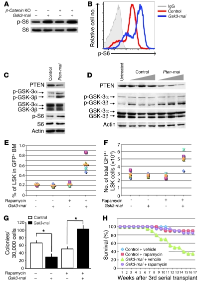
Figure 6. Increased HSCs/HPCs in rapamycin-treated recipients of Gsk3-depleted BM.
(A) BM was harvested from Mx-Cre;β-cateninfl/fl mice treated with or without polyI:polyC, transduced with control or Gsk3-rnai lentivirus, and transplanted into irradiated recipients. After 4 months, GFP+ cells were sorted (pooled from 5 mice per group), and phospho–ribosomal protein S6 (p-S6) was detected in cell lysates by immunoblot. (B) Flow cytometric detection of phospho–ribosomal protein S6 with BM cells in A. (C) Irradiated mice were reconstituted with BM transduced with control vector or Pten-rnai. After 4 months, phospho–GSK-3 and phospho–ribosomal protein S6 were assessed in GFP+ cells by immunoblot. (D) NIH3T3 cells were infected for 3 days with control or Pten-rnai lentivirus, and phospho–GSK-3α/β in control and Pten-depleted cells was detected by immunoblot. (E) Noncompetitive serial transplants. GFP+ cells (2 × 10⁶) from primary recipients of control or Gsk3-rnai were transplanted into 10 irradiated recipients per group. After 1.5–2 months, secondary recipients were injected with rapamycin or vehicle every other day for 8 weeks. Percent GFP+ LSK cells was compared among the 4 groups. (F) Absolute number of GFP+ LSK cells as in E. (G) Colony formation with sorted GFP+ cells from E. (H) Kaplan-Meier plot showing survival of tertiary recipients transplanted with BM from control- and rapamycin-treated secondary recipients in E–G. Shown is control vector BM from secondary recipients treated with vehicle or rapamycin and Gsk3-rnai–infected BM from secondary recipients treated with vehicle or rapamycin transplanted to lethally irradiated tertiary recipients. *P < 0.05.

