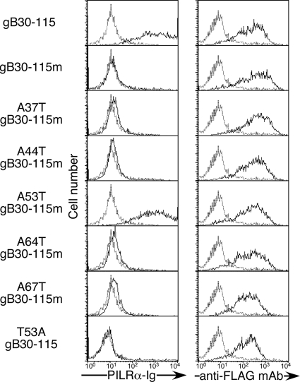
FIG. 2.
Mutational analyses of O glycosylation sites in the N terminus domain of gB. Flag-tagged N terminus fragments of gB (amino acid residues 30 to 115) containing five potential O glycosylation sites or point mutations of these possible O glycosylation sites were transfected into 293T cells. The transfectants were stained with control Ig (dotted line) or PILRα-Ig (solid line). Expression of the N terminus domain of gB was analyzed by staining with anti-Flag MAb (solid line) or control MAb (dotted line). Histograms show fluorescence intensity measured in arbitrary units on a log scale (x axis) and relative cell number on a linear scale (y axis).

