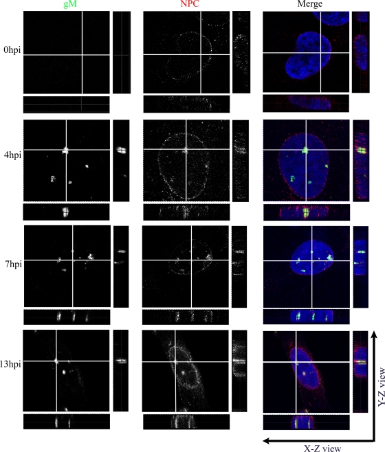
FIG. 2.
gM localizes primarily in punctate extensions and invaginations of nuclear membranes in infected cells. 143B cells were infected with wild-type HSV-1 strain 17+ at an MOI of 2. At the indicated time points, the cells were fixed and stained for gM (green), the NPC using MAb 414 (red), and the nucleus using Topro-3 (blue). The cells were then examined by laser scanning confocal microscopy. The individual color channels were scanned sequentially with only the fluorescence-stimulating laser powered on. Orthogonal slices in the X-Z and Y-Z planes were constructed in which the gray lines represent the cutting positions for the analysis.

