FIG. 1.
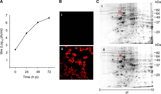
Proteomic analysis of DENV-infected K562 cells. K562 cells were mock (i) or DENV (ii) infected at an MOI of 5. (A) Supernatant was sampled at the indicated time point p.i. and assayed for infectious-virus release by plaque assay. (B) At 72 h p.i., cells were fixed and immunostained for mouse anti-DENV and visualized with goat anti-mouse Alexa546 and confocal microscopy. (C) At 72 h p.i., cells were lysed, the lysates were subjected to 2DGE, and the proteins were visualized with Coomassie blue staining. The boxed protein spot was upregulated in DENV-infected cell lysates and was identified by MS analysis as GRP78. The results are representative of three independent infections.

