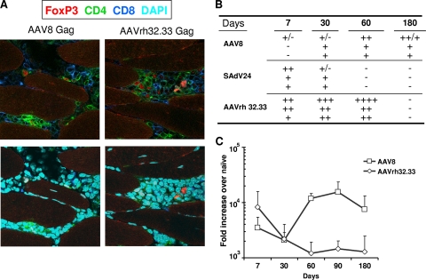
FIG. 4.
Histopathology and Gag antigen expression levels around injection site. Mice were i.m. immunized with 3 × 10¹⁰ GC of AAV8 or AAVrh32.33 expressing HIV Gag. At day 7, 30, 60, 90, and 180 postimmunization, muscle tissue from the injected areas was obtained and analyzed for inflammation, infiltration, and Gag mRNA levels. (A) At 60 days post-vector administration, infiltrated cells were immunophenotyped with antibodies against CD8+ (dark blue), CD4+ (green), and FoxP3 (red). The bottom panels show additional staining with DAPI (light blue). (B) Table scoring the degree of infiltration at different time points and compared with SAdV24 as positive control (−, no infiltrates; ++++, strongest infiltration observed). Data for individual mice in each group are shown. (C) Gag mRNA levels from muscle injected with AAV8 or AAVrh32.33 by TaqMan. Data are shown as mean results with standard deviations.

