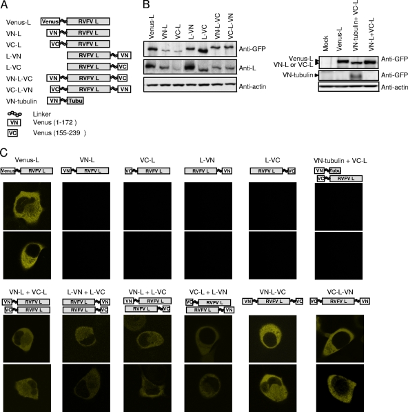
FIG. 6.
BiFC analysis of various full-length L proteins. (A) A schematic diagram of L protein carrying Venus protein at the N terminus (Venus-L) or L proteins carrying an N-terminal fragment of Venus (VN) or a C-terminal fragment of Venus (VC) at their N and/or C terminus with a linker. A schematic diagram of VN-tubulin is also shown. (B) BHK/T7-9 cells in a 12-well plate were independently transfected with 1.0 μg of plasmid expressing the indicated protein. At 12 h posttransfection, intracellular proteins were analyzed by Western blotting by using anti-GFP, anti-L 434, and anti-actin antibodies. (C) BHK/T7-9 cells in a two-well slide were transfected with 1.0 μg of a plasmid or cotransfected with 1.0 μg each of two plasmids as indicated. At 12 h posttransfection, fluorescence in the live cells was observed by confocal microscopy by the use of a YFP filter. For each group, two representative samples are shown.

