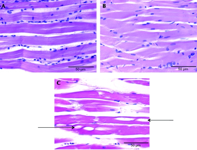
Figure 3.
Photomicrographs of zebrafish musculature. (A) Representative sample of muscle from a zebrafish euthanized with MS222. (B) Representative sample of muscle from a zebrafish euthanized by rapid cooling. (C) Representative sample of muscle from a zebrafish euthanized by rapid cooling and then placed in a –20 °C (–4 °F) freezer. Only (C) had evidence of ice crystal formation (arrows) in the muscle. Hematoxylin and eosin stain; magnification, ×600; bar, 50 µm.

