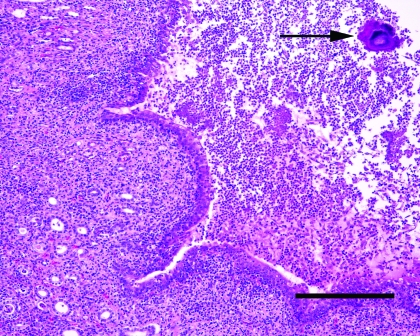
Figure 2.
Kidney from the rat pictured in Figure 1. Note expansion of the renal pelvis by large numbers of degenerate neutrophils and fibrin. Large colonies of bacteria are visible within the exudate (arrow). The transitional epithelium lining the pelvis shows mild squamous metaplasia. Inflammatory cells are also visible within surrounding tubules confirming the presence of a pyelonephritis. Hematoxylin and eosin stain; bar, 200 μm.

