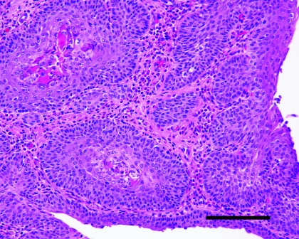
Figure 5.
Ureter from a vitamin-A–deficient rat that did not show clinical evidence of disease. Note the marked squamous metaplasia of the transitional epithelium. Keratin is visible within the epithelium, and moderate numbers of inflammatory cells are present. Hematoxylin and eosin stain; bar, 50 μm.

