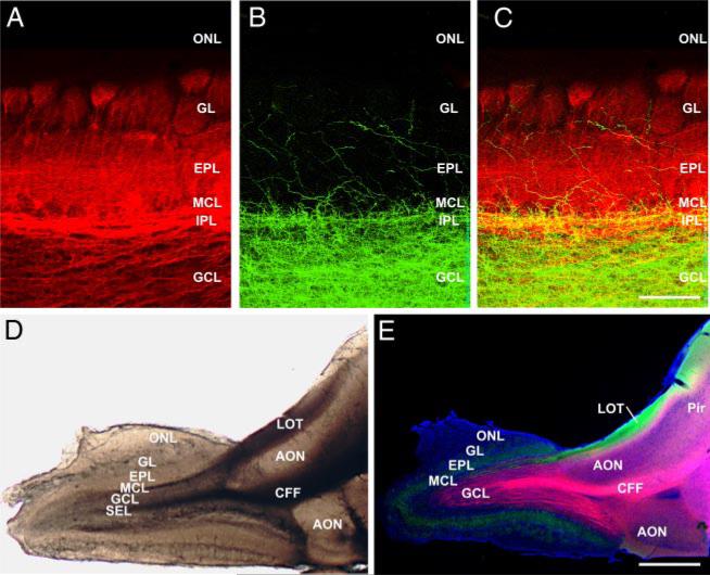
FIG. 1.
Wet mount and tract tracing. A–C: labeling in main olfactory bulb (MOB) after dye injections in vivo. A: retrograde labeling after DiI placement in the lateral olfactory tract (LOT). Mitral/tufted cell axons, soma and dendrites are heavily labeled. B: same section as in A showing anterograde labeling after DiA placement in the centrifugal fiber (CFF) tract. The labeled axons terminate heavily in the granule cell and internal plexiform layers, and more sparsely in the superficial layers. C: overlay image of A and B showing the laminar segregation of labeled fibers and cell bodies. D: wet mount preparation of a 400-μm-thick quasi-horizontal forebrain-MOB slice. The LOT and CFF tract are clearly visible at the level of the anterior olfactory nucleus, just caudal to the MOB. Lateral is at the top, rostral to the left. E: similar slice as in D, in which DiI was placed in the CFF tract and DiA in the LOT; DAPI counterstaining (blue). CFFs (pink staining) project densely into the central bulb while retrograde labeling from the LOT (green staining) distributes through the mitral, external plexiform and glomerular layers. Scale bar = 200 μm in A–C and 1 mm in D and E. AON, anterior olfactory nucleus; EPL, external plexiform layer; GCL, granule cell layer; GL, glomerular layer; IPL, internal plexiform layer; MCL, mitral cell layer; ONL, olfactory nerve layer; Pir, piriform cortex; SEL, subependymal layer.

