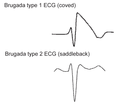
Figure 3).
The two predominant electrocardiogram (ECG) repolarization abnormalities observed in the Brugada syndrome. Brugada syndrome ECG pattern abnormalities are observed in the anterior precordial leads (V1 and/or V2). ECG abnormalities may be dynamic, and alternate between normal or abnormal ECG patterns. The coved-shaped pattern is suggested to be associated with an increased risk of cardiac events

