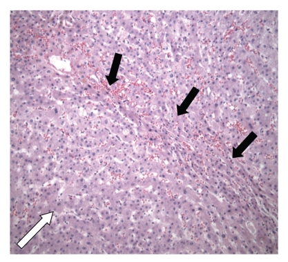
Figure 3.
Detail of the intra-operative liver specimen (Case 2) showing nodular regenerative hyperplasia in which a regenerative nodule (white arrow) is bordered by irregular aligned small-sized hepatic trabeculae (black arrows). Hematoxylin and eosin, original magnification 100x.

