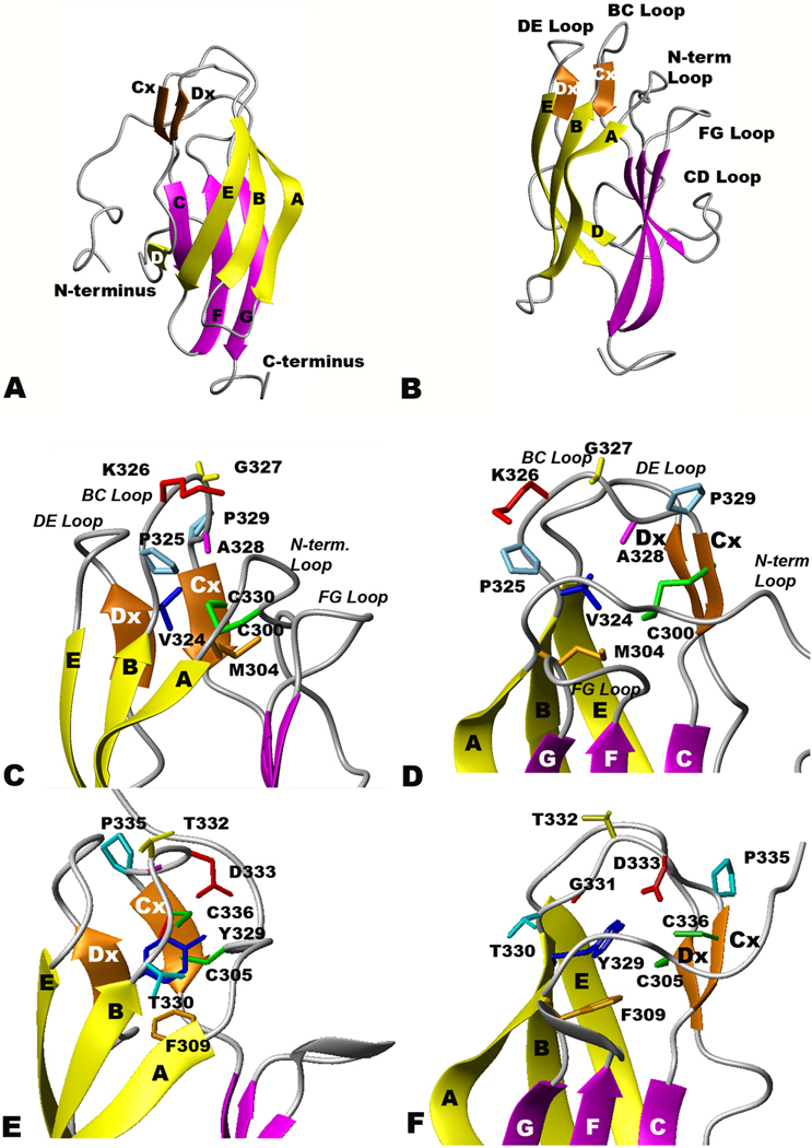
Figure 1.

Ribbon diagrams of rED3 of YFV and WNV. Beta sheets 1–3 are colored yellow, orange and magenta, respectively, and the disulfide bridge between C300 and C330 is colored green. (A and B) Two orthogonal views of the NMR-derived YF rED3 backbone atom structures (C and D) Surface loop structure of YF rED3, and (E and F)WN rED3 (Volk et al., 2004).
