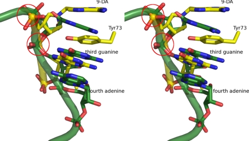
Fig. 5.
Stereo-view of the superimposed inhibitors bound to SAP (yellow, PDB ID code 3HIW) and an unbound GAGA tetraloop (green, PDB ID code 1Q9A). The partial nucleic acid backbone of the inhibitor (yellow) and GAGA tetraloop (green) is shown. Tyr-73 and 9-DA and the third guanosine are depicted in yellow. The last 3 nucleotides of the unbound GAGA tetraloop are shown in green. The 5′- and 3′-phosphate diesters of 9-DA are highlighted in the red circles.

