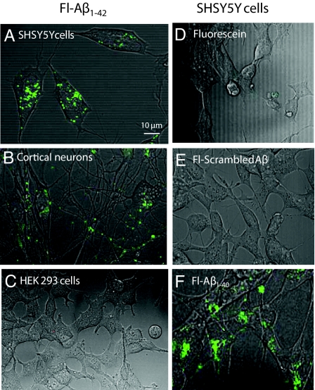
Fig. 1.
Cell uptake of FITC-Aβ. (A) SHSY5Y cells, (B) primary murine cortical neurons, and (C) HEK293 cells were cultured in the presence of 250 nM human FITC-Aβ1–42 for 24 h, then imaged with confocal/phase-contrast microscopy. Vesicular uptake was observed only in the neurons and SHSY5Y cells. (D) SHSY5Y cells were incubated with 250 nM fluorescein alone, (E) FITC-scrambled Aβ1–42, or (F) FITC-Aβ1–40 for 24 h. Vesicular uptake was observed with FITC-Aβ1–42 and Aβ1–40, but not with FITC-scrambled-Aβ1–42 or fluorescein alone.

