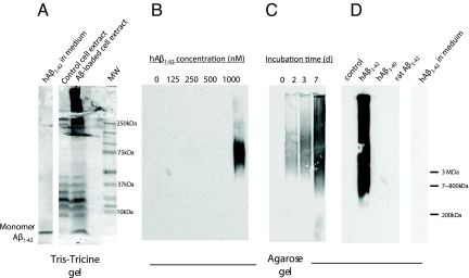
Fig. 5.
Aggregation of intracellular Aβ1–42 into HMW forms. (A) SHSY5Y cells, grown in the presence or absence of Aβ1–42 (1 μM) for 5 days, were homogenized and run on a Tris-Tricine gel and blotted with an anti-Aβ antibody (6E10). Culture medium incubated with Aβ1–42 (1 μM) for 5 days show the presence of only monomers. Extracts from cells grown in Aβ1–42 show HMW aggregates, while cell grown without Aβ do not (note the non-specific bands in the 10–40 kDa range from the cell extracts). (B–D) SHSY5Y cells were grown in the presence of Aβ1–42 (0–1,000 nM as indicated) for 5 days (B), for varying times (0–7 days, 1 μM Aβ) (C), or in the presence of human Aβ1–42, Aβ1–40, or rat Aβ1–42 for 5 days (as indicated in D). Cell homogenates were then run on an agarose gel and blotted with an anti-Aβ antibody (6E10). Intracellular Aβ was found to aggregate in a concentration- and time-dependent manner. Human Aβ1–42 forms HMW intracellular aggregates, but human Aβ1–40 and rat Aβ1–42 do not. Human Aβ1–42 incubated in culture medium alone for 5 days did not form HMW aggregates (D, right lane).

