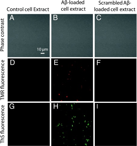
Fig. 6.
Cell extracts from Aβ1–42-loaded cells seed the formation of amyloid fibrils. SHSY5Y cells incubated with or without 1 μM unlabeled Aβ1–42 or scrambled Aβ1–42 for 5 days were homogenized and then incubated with 100 nM TMR-Aβ1–42 for 48 h. Phase contrast images of the cell extracts show the absence of cells (A–C). Extracts from control cells (grown in the absence of Aβ) did not show TMR fluorescence (D) or Thioflavin-S staining (G); Aβ-loaded cell extracts developed TMR precipitates (E) which stained for Thioflavin-S (H); while scrambled-Aβ-loaded cell extracts showed neither (F and I). These results suggest that intracellular Aβ aggregates can seed the formation of Thioflavin-positive aggregates.

