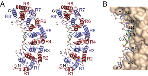
Fig. 1.
Crystal structure of FBF-2 in complex with gld-1 FBEa RNA. (A) Stereo-view of FBF-2 in complex with gld-1 FBEa RNA. Repeats are colored alternately red and blue. Side chains that interact with RNA are shown. The RNA is colored by atom type (gray, carbon; red, oxygen; blue, nitrogen; orange, phosphorus; yellow, sulfur). Bases 4–6 are shown with green carbon atoms. (B) Surface representation of FBF-2 in complex with gld-1 FBEa RNA. The figures were prepared with PyMol (44).

