Figure 2.

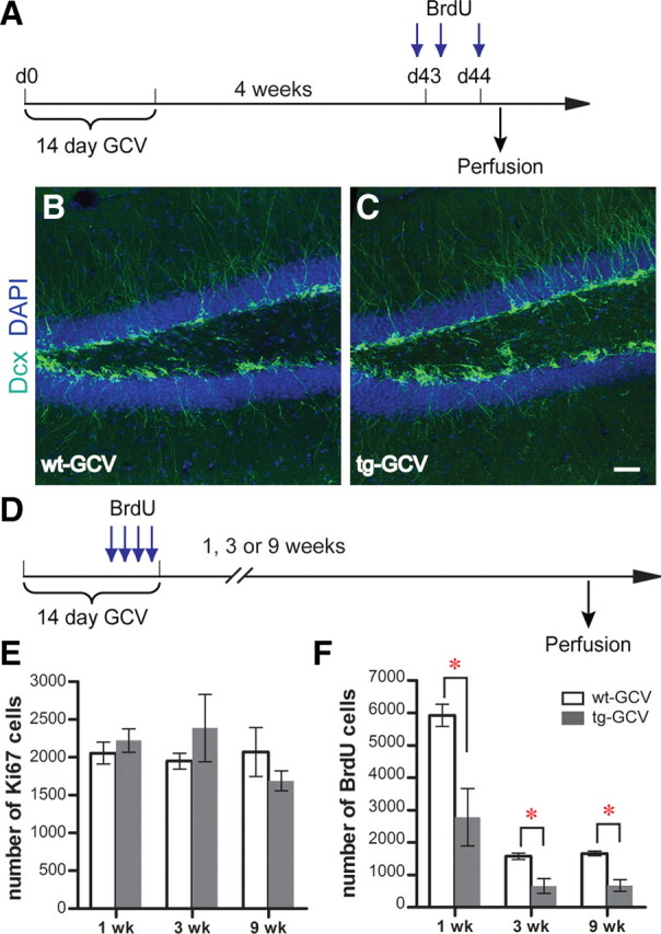
Recovery of progenitor cell proliferation and neurogenesis in the DG after drug withdrawal in Nestin-tk transgenic mice. A, The experimental scheme for B and C. B, C, Representative confocal images of adult DG from wt-GCV and tg-GCV mice labeled by immature neuron marker Dcx (green). Nuclear marker DAPI is in blue. See text for quantifications. D, The experimental scheme for E and F. E, Ki67 cell numbers are similar between tg-GCV and wt-GCV at 1, 3, and 9 weeks after GCV treatment. F, BrdU cell numbers are reduced in tg-GCV at 1, 3, and 9 weeks after GCV treatment. See supplemental Table 1, available at www.jneurosci.org as supplemental material, for statistics for E and F. Scale bar: (in C) B and C, 50 μm. *p < 0.01. Error bars represent ± SEM.
