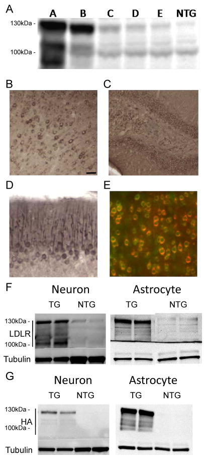
Figure 1. Expression of LDLR Transgene in Neurons and Astrocytes.
(A) Levels of LDLR protein in the cortex of 5 different LDLR transgenic lines were assessed by western blotting. RIPA-soluble cortex lysates from LDLR transgenic mice and non-transgenic (NTG) mice were probed with anti-LDLR antibody (Novus). (B–D) Regional expression patterns of LDLR in B line mice were characterized by immunostaining with anti-HA antibody to detect HA-tagged LDLR protein. LDLR was expressed in the cortex (B), dentate gyrus of hippocampus (C), and Purkinje cell dendrites of cerebellum (D). (E–G) Cellular expression profile of the LDLR transgene was examined by using anti-HA or anti-LDLR antibody. (E) Cortical sections were stained by double-immunofluorescence labeling for HA (red) and the neuronal marker NeuN (green). (F) Cell lysates from primary neurons or astrocytes isolated from LDLR B line transgenic (TG) and NTG mice were analyzed by probing with either anti-LDLR (Novus) or anti-LDLR (Dr. Bu) antibody, respectively. (G) Expression of HA-tagged LDLR transgene in primary neurons and astrocytes was confirmed by western blotting with anti-HA antibody. Scale bar: 30μm. See also Figure S1.

