Figure 2.
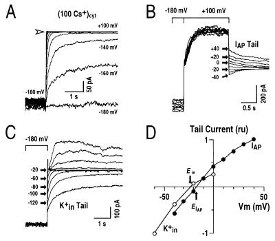
Transient outward currents are carried mainly by K+. (A) Whole-cell current recordings under conditions with 100 mM Cs+ in the pipette solution. The voltage protocol was similar to that given in Fig. 1B (note large K+in currents at potentials negative of −100 mV). Arrow shows zero current level. (B) Tail current recordings of the transient outward currents. From a holding potential of −180 mV, transient outward currents were activated by a voltage step to +100 mV. Tail currents of the transient outward currents were recorded at the indicated membrane potentials (IAP Tail). (C) Tail currents of K+in currents recorded in the same cell as in B. After a prepulse potential of −180 mV, where K+in currents were activated, tail currents were recorded at the indicated membrane potentials ranging from −140 mV to +80 mV with +20 mV increments (K+in Tail). At increasingly depolarized tail potentials, the decay times of K+in current deactivation became shorter and therefore could be clearly distinguished from the transient outward currents. (D) Tail currents from recordings in B and C were plotted as a function of applied membrane potentials. ru, relative units. IAP tail currents and K+in tail currents were normalized by the currents at +40 mV and −60 mV, respectively. Pipette solutions with zero [Ca2+]i and the standard bath solution were used.

