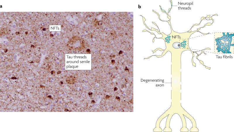
Figure 1. Tau pathology in AD and related tauopathies.
At autopsy, the brains of patients with Alzheimer's disease or related tauopathies show abundant neurofibrillary tangles (NFTs) and neuropil threads that are comprised of pathological tau. These tau deposits can be visualized by treating brain slices with certain silver stains or by immunostaining with antibodies that recognize tau (as shown in A, with darkly-stained NFTs and dense tau neuropil threads that yield a nearly uniform brown staining in a hippocampal section of an Alzheimer's disease brain). A schematic representation of NFTs and neuropil threads within a neuron is shown in B, with an example of tau fibrils that resemble those found in NFTs depicted in the associated inset.

