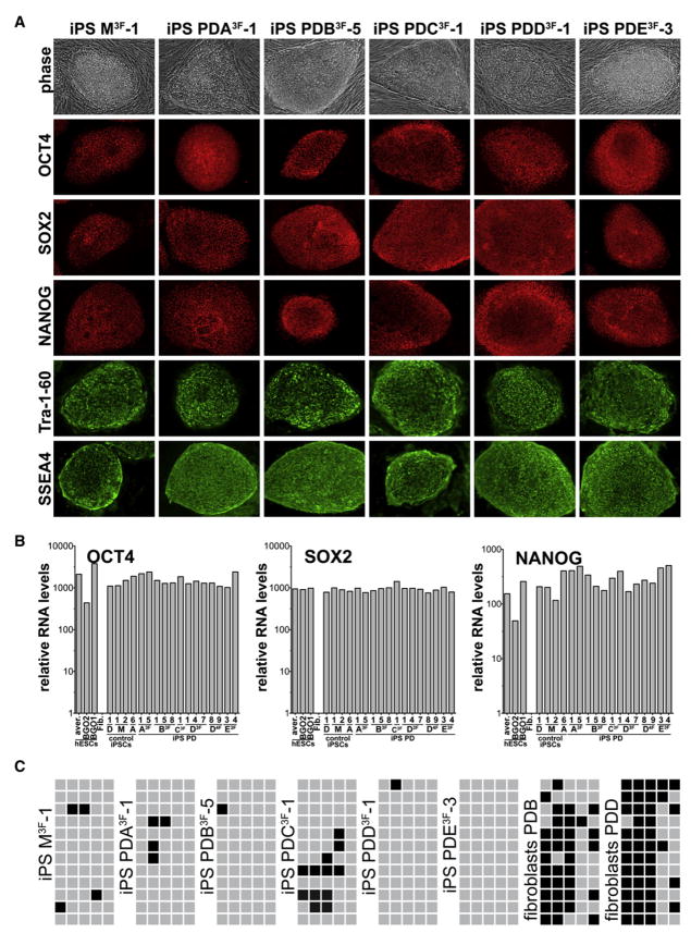
Figure 1. Characterization of DOX-Inducible hiPSCs Derived from Fibroblasts from PD Patients.
(A) Phase contrast picture and immunofluorescence staining of hiPSC lines M3F-1 (non-PD hiPSCs), PDA3F-1, PDB3F-5, PDC3F-1, PDD3F-1, and PDE3F-3 for pluripotency markers SSEA4, Tra-1-60, OCT4, SOX2, and NANOG.
(B) Quantitative RT-PCR for the reactivation of the endogenous pluripotency-related genes NANOG, OCT4, and SOX2 in indicated hiPSC lines, hESCs, and primary fibroblasts. Relative expression levels were normalized to expression of these genes in fibroblasts.
(C) Methylation analysis of the OCT4 promoter region. Light gray squares indicate unmethylated and black squares methylated CpGs in the OCT4 promoter of hiPSCs and parental primary fibroblasts cells.

