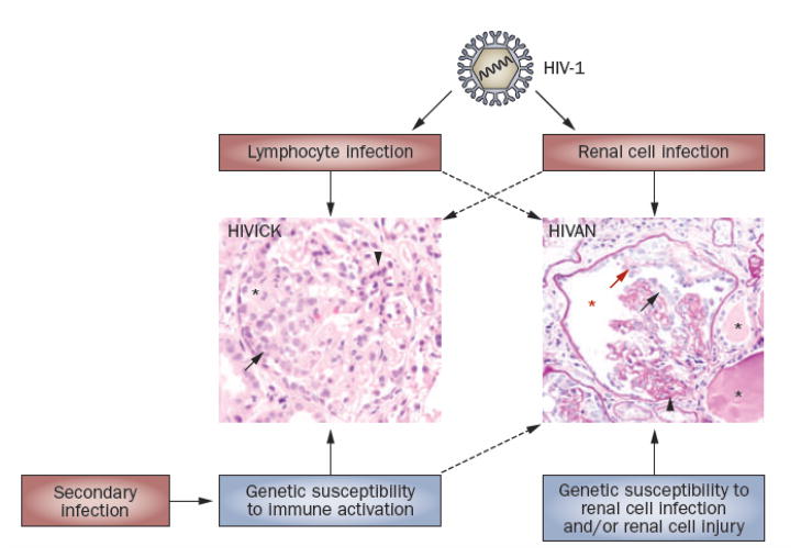
Figure 2.
The interplay of environmental (red) and genetic (blue) factors in HIV-associated renal diseases. Of central importance is HIV-1 infection, which occurs in both renal epithelial cells and immunocytes resident and infiltrating the kidney. Although their effect is not fully understood, genetic factors also contribute to pathogenesis, possibly by determining susceptibility to renal cell infection or cell injury response. In HIVICK, and perhaps also HIVAN, immune and inflammatory pathways have a role in disease onset, progression, and severity. Top panel shows typical HIVAN pathology consisting of collapse of the glomerular tuft with segmental areas of sclerosis (thickened basement membranes) and proliferation of epithelial cells adherent to both the tuft and Bowman’s capsule. Microcysts with proteinacious casts can be seen in adjacent tubules. Bottom panel is typical pathology of HIVICK with global sclerosis, mesangial expansion, and hypercellularity encompassing the tuft and Bowman’s capsule. For more detailed information on the pathology of these HIV-associated renal disease, please see an accompanying article in the series. Hematoxylin and eosin stain, both images 60X magnification. Abbreviations: HIVAN, HIV-associated nephropathy; HIVICK, HIV-immune-complex kidney disease..

