
Figure 3.
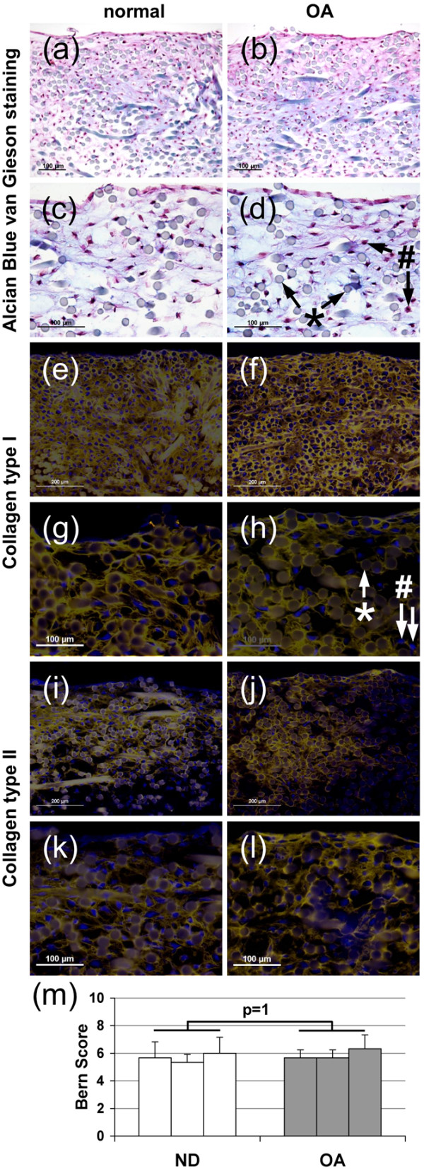
Histology of osteoarthritic and normal chondrocyte scaffold culture. Chondrogenic differentiation of chondrocytes obtained from (a, c, e, g, i, k) normal and (b, d, f, h, j, l) osteoarthritic (OA) articular cartilage cultured in Hyaff-11 scaffolds. (a to d) Alcian Blue van Gieson staining, immunohistochemical localization of collagen (e to h) type I and (I to l) type II, with (g, h, k, l) higher magnification, and (m) Bern Score, * scaffold fibre, # cell nuclei. Three cultures per donor group.