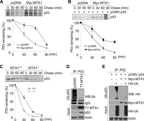
FIGURE 3.
MTA1 regulates p53 protein stability by inhibiting its ubiquitination. A and B, U2OS (A) or H1299 (B) cells were transfected with the indicated expression vectors. After 36 h of transfection, cells were labeled using [35S]methionine and subjected to pulse chase analysis. Cells were harvested at various time points during the chase period and immunoprecipitated using an anti-p53 antibody. Complexes were resolved by SDS-PAGE and exposed to storage phosphor screens. The intensity of the labeled p53 band was quantified by phosphorimage analysis using ImageQuant software (Molecular Dynamics), and the percent p53 remaining was calculated relative to that at the beginning of the chase period (time 0). The mean values from three independent experiments are shown. C, MTA1+/+ and MTA1−/− MEFs were labeled using [35S]methionine and subjected to pulse chase analysis as described above. D, protein extracts from the MCF-7/pcDNA and MCF-7/T7-MTA1 cells were subjected to IP with an anti-p53 antibody, following by Western blot analysis with the indicated antibodies. E, HEK293 cells were transfected with the indicated plasmids and subjected to the sequential IP/Western blot analysis as described above.

