FIGURE 5.
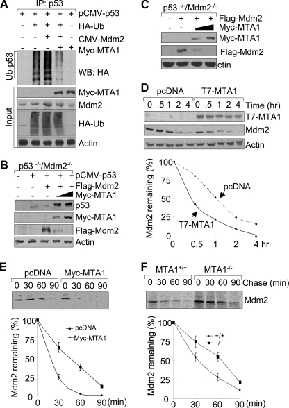
MTA1 inhibits the Mdm2-mediated degradation of p53. A, HEK293 cells were transfected with the indicated plasmids and subjected to the sequential IP/Western blot analysis as described above. B and C, p53−/−/Mdm2−/− double-null MEFs were transfected with the indicated expression vectors and immunoblotted with the indicated antibodies. D, MCF-7/pcDNA and MCF-7/T7-MTA1 cells were treated with 100 μg/ml of cycloheximide and harvested at indicated times for Western blot analysis with the indicated antibodies (upper panel). Western blots were subjected to densitometric analysis and results were normalized based on actin expression levels and reported in graphical form (lower panel). E and F, U2OS cells transfected the indicated expression vectors (E) or MTA1+/+ and MTA1−/− MEFs (F) were labeled using [35S]methionine and subjected to pulse chase analysis as described above except that immunoprecipitations were carried out using an anti-Mdm2 antibody.

